Periodontitis is an inflammation of the connective tissue (periodontium) located between the dental root cement and the alveolar plate (bone cavity). Infection that develops in the root canals, through the apical opening, spreads beyond the borders of the root apex to the surrounding tissues and structures. It can be acute or chronic. Treatment of periodontitis includes the elimination of infection in the dental canals, their thorough cleaning and filling.

Causes
Periodontitis of a tooth is usually a complication of a cavity, pulpitis, or infection of the dental canal due to a broken filling. It can also be caused by a fracture, root trauma or overloading of the dental unit due to improper prosthetics. Periodontal inflammation can be caused by bad habits such as holding a pencil, pen, pipe, or a filling placed above the bite that regularly exerts abnormal pressure on the tooth. Sometimes periodontitis develops after treatment of a tooth due to strong medications getting into the periodontium.
Types of periodontitis
Periodontal inflammation can be acute (serous, purulent) and chronic (fibrous, granulomatous, granulomatous). Periodontitis is subdivided by localization into apical and marginal, and by cause into infectious, traumatic and medication-associated.
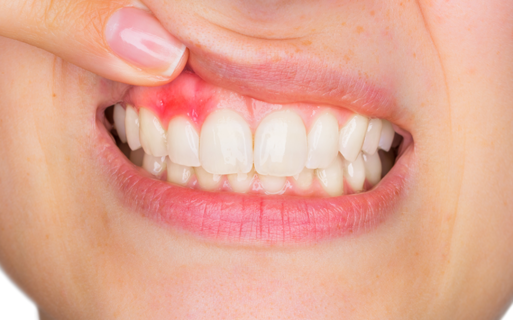
The acute form is manifested by pain when loading the tooth, which grows and becomes intolerable as the pathological process spreads. There is a feeling of oppression in the tooth and it is impossible to touch it. Often there is swelling and reddening of the gum, fistulas and pus may develop. The tooth changes color and becomes mobile (with destruction of the periodontal ligament).
The chronic form is almost asymptomatic. When the chronic process is exacerbated, there are pronounced reactions – intense pain, the sensation of the tooth tearing from the inside. An asymptomatic course of the disease is dangerous, because the inflammation spreads further to healthy tissues. Such a condition is dangerous with the formation of cysts, granulomas, osteomyelitis, and endogenous maxillary sinusitis.
Chronic periodontitis is a mine with delayed action. The infectious focus serves as a trigger for the development of an acute purulent process. The purulent form is often accompanied by the formation of an endogenous fistula with the release of purulent contents to the outside, or more dangerous consequences – abscess, phlegmon, up to sepsis.
Signs of acute periodontitis
- Constant nagging pain.
- Severe pain when straining the tooth.
- Increased sensitivity to temperature, chemical stimuli.
- A clear understanding of exactly which tooth hurts (a feeling of distension from the inside).
- Pain may be nagging, pulsating, or may extend to the ear or temple.
- There is a foul odor in the mouth.
- Swelling and redness of the gum.
- Pus discharge, fistula formation.
- Moveability of the tooth (if the periosteum is destroyed).
Diagnosis
The success of treatment depends on the accuracy of diagnosis and the quality of cleaning and sterilization of the intracanal system of the tooth. Missing even one micro branch of the dental canal is fraught with serious complications. To determine the nature, extent, and form of periodontitis of the tooth, a comprehensive diagnosis is performed, including:
- Targeted imaging of the affected tooth.
- Computer tomography to obtain a three-dimensional image.
- Instrumental examination under a microscope.
- Complex approach allows to study the pathological focus, structure of the root system in detail, determine the number of dental canals, assess their condition, localization and size of the inflamed area. Examination under a microscope allows the dentist to examine the root canals in detail, including all their branches, to detect microcracks and other defects.
Methods of treatment
The treatment of periodontitis is aimed at solving several problems:
- Elimination of the pathological process, prevention of further spread of inflammation.
- Preservation and restoration of the anatomy of the affected unit and the functionality of the entire dentoalveolar apparatus.
The general approach to treatment includes:
- Creation of access to the dental cavity and its opening.
- Creation of access to dental canals.
- Passage of canals, their unsealing (if the tooth was previously pulped and filled), determining the length with an apex locator.
- Mechanical and medicinal treatment of the root canals (removal of diseased tissue to healthy boundaries, antiseptic treatment for disinfection).
- Temporary filling – for the complete destruction of the infection, a drug is put into the root canal, closed from above with a temporary filling. Under the influence of the drug, pathogenic microflora dies, strengthens the walls of the tooth and the bone of the hole.
- A permanent filling – after 2-3 weeks, the drug is removed, the condition of the peri-tooth tissues is evaluated using X-ray examination. If there is no infectious inflammatory process, the canals are sealed hermetically, without allowing the formation of air voids.
- Restoration after endodontic treatment – depending on the degree of destruction of the crown portion of the tooth, artistic restoration (restoring the anatomic shape using modern nano-polymers) or inlay prosthetics, crowns are used. Orthopedic structures are used when the supragingival part of the tooth is damaged by 50% or more.
If conservative therapy is inefficient or inappropriate, the doctor considers surgical treatment:
- Apex resection of the tooth (apicoectomy) – excision of the injured part of the root along with the pathologically altered tissues.
- Coronary-radicular separation – separation of the roots by dissecting the stem and crown of the tooth. It is used on 2-rooted teeth if the focus of inflammation is located in the area of root divergence.
- Hemisection of the tooth – amputation of the affected root in multi-rooted units along with the adjacent crown.
- Removal of the tooth unit with subsequent implantation and prosthetics.
The method of treatment is chosen according to the clinical picture. The choice of intervention tactics is influenced by:
- Features of the anatomy of the tooth – too curved, intertwined roots will not allow for quality cleaning, disinfection of the canals.
- Condition of the tooth unit – the absence or presence of root canal obliteration, fractures, root perforation, the presence of inlays, pins, which cannot be removed without severe damage to the root, etc.
- The presence of concomitant pathologies (multiple root cysts, granulomas).
- The possibility to isolate the working field qualitatively, to provide full access to the dental canals.
If the treatment of periodontitis is carried out qualitatively, there will be no recurrence.
