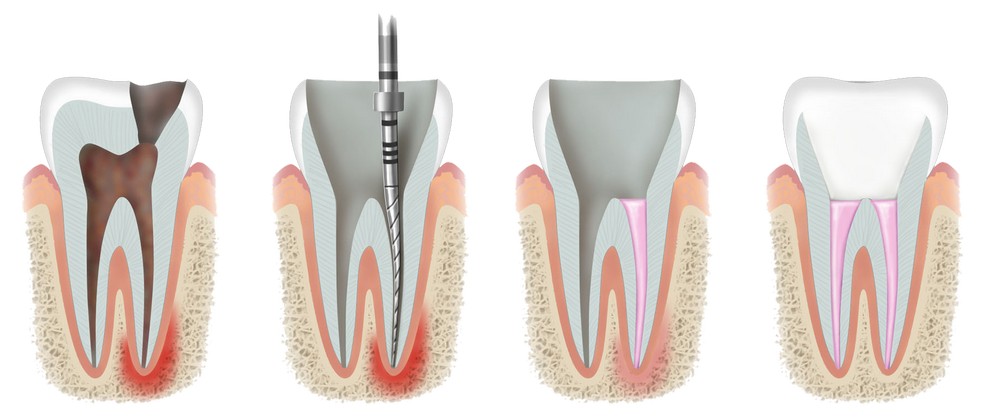
The root canal is the part of the tooth that contains the pulp, or dental nerve. The soft fibrous pulp consists of connective tissue, nerve endings and blood vessels. A “living” tooth differs from a “dead” one precisely in the presence of pulp, which nourishes the tooth tissues.
Often, problems that arise during endodontic treatment are associated with a lack of medical knowledge about the anatomical and morphological features of root canals. But this is a fundamentally important point that affects the quality of work.
Canals in structure in different groups of teeth differ from each other. Teeth can have both main and lateral, additional canals located at different levels and having a simple or complex configuration. Dental canals can be compared to the roots of a tree – if you look closely at them, it becomes obvious that their shape is unique.
It should be emphasized that lateral canals located in any part of the root are found in more than 50% of cases. Often additional canals branch at an angle to each other, end blindly, communicate with the periodontium through additional branches or through the main canal. All this creates difficulties in treatment. The dentist must be familiar not only with the variants of the norm, but also with deviations in the direction and shape of the dental passages.
It is customary to distinguish 9 configurations of dental canals, although this scheme can be considered approximate – in reality, there are many more variations. This phenomenon is associated not only with genetic factors, but also with phenotypic (developed in the course of life) changes. Thus, the high frequency of the appearance of single-rooted teeth with two canals is often due to the process of fusion of individual roots.
Root Canal Diseases
The main endodontic problems are pulpitis and periodontitis. They develop as complications of advanced caries, as well as periodontal diseases, with gum or tooth injuries. The infectious process causes the patient not only moral and physical discomfort, but also causes odontogenic inflammation of the neck and maxillofacial region.
The main stages of treatment
pulp removal
cleaning of the pulp chamber and dental canals
final stroke formation
filling
Endodontic treatment methods should be aimed at saving even the most damaged and infected teeth, at preventing the occurrence of inflammation of bone and soft tissues leading to tooth loss. In addition, it is important to restore the anatomical and physiological function of the damaged dental unit with the help of filling material.
Features of the treatment of teeth with thin, crooked canals
A competent preliminary examination in this case is a paramount task. The shape of the crooked canal, as a rule, changes along its entire length, the lumen is not round, but slit-like. This is important to consider when processing – all loops and side branches must be thoroughly cleaned and securely sealed. The preparation of thin canals should be carried out with great care – the probability of perforation of the root canal is high.
To identify the structural features of the dental canals, targeted radiography, computed tomography, an apex locator and a dental microscope are used.
After determining the number, length and condition of the channels, they are instrumented. This is one of the most difficult steps. The main goal is to thoroughly clean the channel and give it a cone shape. To work with crooked canals, ultra-fine instruments (tip diameter 0.06 mm) are used. The desired shape is created in two ways:
manual
machine, using an endodontic handpiece
The second method in the treatment of thin canals is preferable, because. allows you to achieve perfect smoothness of the walls, which greatly facilitates further work. Washing problem channels should be carried out repeatedly to remove the microbial film completely. The final stage is filling with gutta-percha in combination with various sealants and plastics.
What is a root canal revision?
Revision (retreatment) differs from primary endodontic intervention in the difficulty of processing due to the need to eliminate foreign bodies and the material with which the canals were sealed earlier. Often you have to remove orthopedic structures or replace them. In addition, revision is an additional stress for the patient.
That is why in the past, many patients, despite the need for revision, made a choice in favor of surgical intervention or completely let everything take its course, leaving it as it is. Today, with the advent of depophoresis, the situation has changed. Depophoresis is a modern technique for treating dental canals using copper-calcium hydroxide and a weak electric field. It becomes indispensable for curved canals with numerous perforations in them.
The use of heated gutta-percha in the treatment of crooked and thin canals makes it possible to seal the passages throughout without voids and minimize the possibility of complications. The use of modern tools and materials, the practical application of the latest achievements of dental science and an individual approach to each client allow successful therapy even in very difficult cases.
